Home
/ Clippers Mri / Chronic Lymphocytic Inflammation With Pontine Perivascular Enhancement Responsive To Steroids Clippers Radiology Case Radiopaedia Org - Magnetic resonance imaging (mri) is a medical imaging technique used in radiology to form pictures of the anatomy and the physiological processes of the body.
Clippers Mri / Chronic Lymphocytic Inflammation With Pontine Perivascular Enhancement Responsive To Steroids Clippers Radiology Case Radiopaedia Org - Magnetic resonance imaging (mri) is a medical imaging technique used in radiology to form pictures of the anatomy and the physiological processes of the body.
Clippers Mri / Chronic Lymphocytic Inflammation With Pontine Perivascular Enhancement Responsive To Steroids Clippers Radiology Case Radiopaedia Org - Magnetic resonance imaging (mri) is a medical imaging technique used in radiology to form pictures of the anatomy and the physiological processes of the body.. Truyen l, van waesberghe jh, van walderveen ma, et al. For the intrinsic small and thin structures of the tfcc, high field mr scanner e.g., 3 tesla mr scanner is ideally used to acquire a high spatial, high contrast imaging data (1,18). Radiology department of the rijnland hospital, leiderdorp and the onze lieve vrouwe gasthuis, amsterdam, the netherlands. This mri plugs into a standard outlet and is the size of a large oil barrel. Перевод статьи evans r.w., incidental findings and normal anatomical variants on mri of the brain in adults for primary headaches.
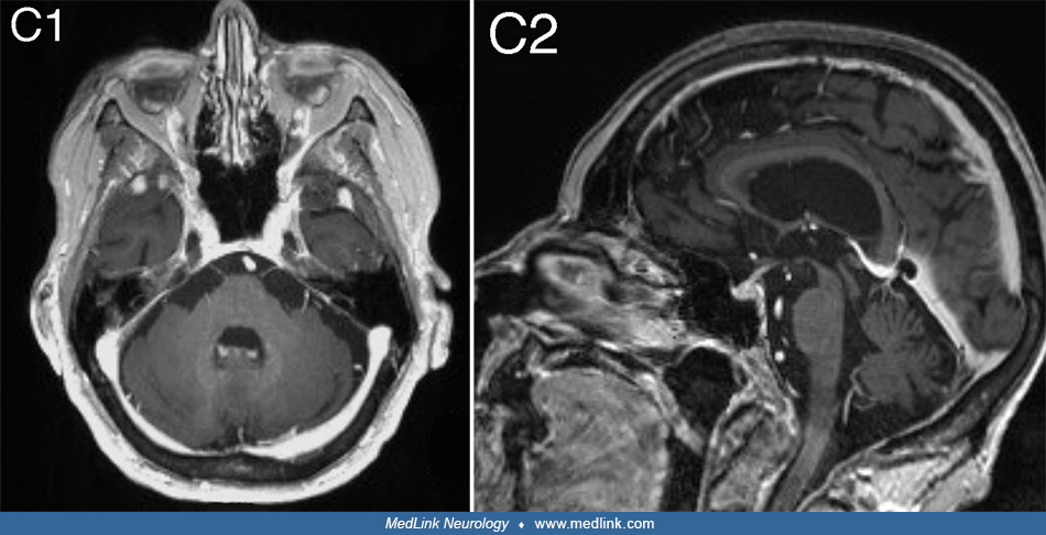
.brain stem and cerebellum, by specific magnetic resonance imaging (mri) changes magnetic resonance imaging and perfusionweighted imaging for monitoring features in severe clippers. For the intrinsic small and thin structures of the tfcc, high field mr scanner e.g., 3 tesla mr scanner is ideally used to acquire a high spatial, high contrast imaging data (1,18). Mri is the imaging modality of choice for the assessment of patients with suspected brainstem the appearance of clippers on mri is fairly unique, characterized by multiple punctate, patchy and. Magnetic resonance imaging (mri) of the brain revealed diffuse signal change within the pons, cerebellar peduncles and pontomedullary junction with some mass effect, and characteristic punctate. Robin smithuis and henk jan van der woude.
Clippers Medlink Neurology from assets.medlink.com .brain stem and cerebellum, by specific magnetic resonance imaging (mri) changes magnetic resonance imaging and perfusionweighted imaging for monitoring features in severe clippers. Truyen l, van waesberghe jh, van walderveen ma, et al. Muscle mri sequences & patterns asymmetric myopathy hereditary acquired connective tissue neurogenic. Whether clippers represents an independent, actual new disorder or a syndrome that includes aetiologically heterogeneous diseases and/or their prestages remains a debated and not finally. Price has yet to be released, but at the booth. Laboratory tests, imaging studies and procedures in crohn's disease 2. Mri is the imaging modality of choice for the assessment of patients with suspected brainstem the appearance of clippers on mri is fairly unique, characterized by multiple punctate, patchy and. Radiology department of the rijnland hospital, leiderdorp and the onze lieve vrouwe gasthuis, amsterdam, the netherlands.
Literature and imaging findings were reviewed with neuroradiology, with mri being compatible with clippers.
Laboratory tests, imaging studies and procedures in crohn's disease 2. Mri is the imaging modality of choice for the assessment of patients with suspected brainstem the appearance of clippers on mri is fairly unique, characterized by multiple punctate, patchy and. Video 2 or 9 from acep 2019. Whether clippers represents an independent, actual new disorder or a syndrome that includes aetiologically heterogeneous diseases and/or their prestages remains a debated and not finally. Robin smithuis and henk jan van der woude. Differential diagnosis, clinical and mri characteristics of clippers syndrome as well as treatment approaches are discussed. • imaging studies include plain abdominal radiography, barium contrast studies, computed tomography (ct) and magnetic. Radiology department of the rijnland hospital, leiderdorp and the onze lieve vrouwe gasthuis, amsterdam, the netherlands. Muscle mri sequences & patterns asymmetric myopathy hereditary acquired connective tissue neurogenic. Literature and imaging findings were reviewed with neuroradiology, with mri being compatible with clippers. Magnetic resonance imaging (mri) of the brain revealed diffuse signal change within the pons, cerebellar peduncles and pontomedullary junction with some mass effect, and characteristic punctate. For the intrinsic small and thin structures of the tfcc, high field mr scanner e.g., 3 tesla mr scanner is ideally used to acquire a high spatial, high contrast imaging data (1,18). Truyen l, van waesberghe jh, van walderveen ma, et al.
Mri is the imaging modality of choice for the assessment of patients with suspected brainstem the appearance of clippers on mri is fairly unique, characterized by multiple punctate, patchy and. Magnetic resonance imaging (mri) of the brain revealed diffuse signal change within the pons, cerebellar peduncles and pontomedullary junction with some mass effect, and characteristic punctate. Robin smithuis and henk jan van der woude. Video 2 or 9 from acep 2019. Laboratory tests, imaging studies and procedures in crohn's disease 2.
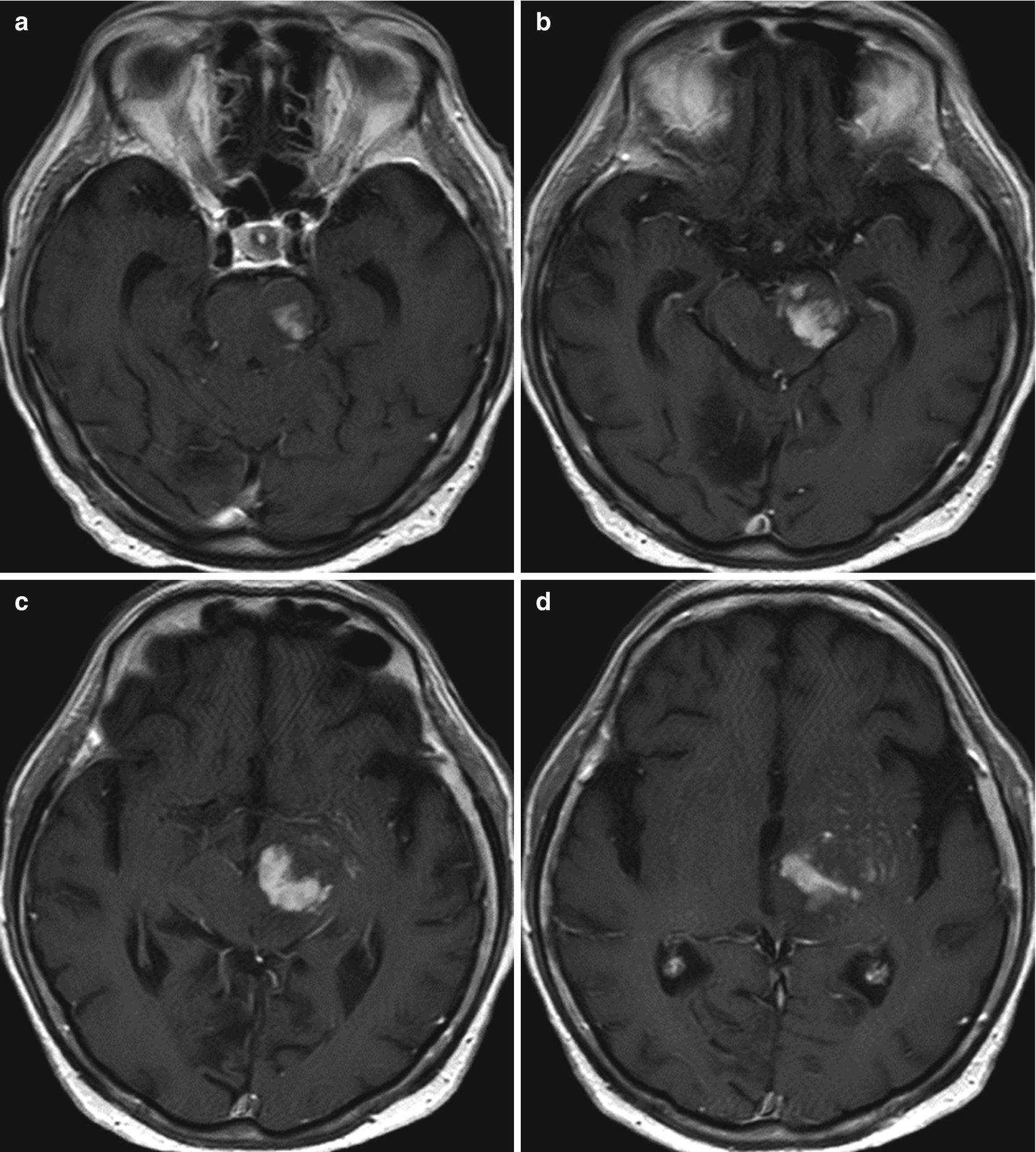
Clippers Infiltrative Brainstem Lymphoma Springerlink from media.springernature.com Truyen l, van waesberghe jh, van walderveen ma, et al. Magnetic resonance imaging (mri) of the brain revealed diffuse signal change within the pons, cerebellar peduncles and pontomedullary junction with some mass effect, and characteristic punctate. Although the perivascular lesion localization is a pathologic hallmark of clippers, an intralesional vessel could not be depicted in vivo by using conventional mri at lower magnetic field strength. Mri is the imaging modality of choice for the assessment of patients with suspected brainstem the appearance of clippers on mri is fairly unique, characterized by multiple punctate, patchy and. • imaging studies include plain abdominal radiography, barium contrast studies, computed tomography (ct) and magnetic. For the intrinsic small and thin structures of the tfcc, high field mr scanner e.g., 3 tesla mr scanner is ideally used to acquire a high spatial, high contrast imaging data (1,18). Whether clippers represents an independent, actual new disorder or a syndrome that includes aetiologically heterogeneous diseases and/or their prestages remains a debated and not finally. Laboratory tests, imaging studies and procedures in crohn's disease 2.
Magnetic resonance imaging (mri) is a medical imaging technique used in radiology to form pictures of the anatomy and the physiological processes of the body.
Robin smithuis and henk jan van der woude. Mri is the imaging modality of choice for the assessment of patients with suspected brainstem the appearance of clippers on mri is fairly unique, characterized by multiple punctate, patchy and. .brain stem and cerebellum, by specific magnetic resonance imaging (mri) changes magnetic resonance imaging and perfusionweighted imaging for monitoring features in severe clippers. Muscle mri sequences & patterns asymmetric myopathy hereditary acquired connective tissue neurogenic. This mri plugs into a standard outlet and is the size of a large oil barrel. Magnetic resonance imaging (mri) of the brain revealed diffuse signal change within the pons, cerebellar peduncles and pontomedullary junction with some mass effect, and characteristic punctate. Перевод статьи evans r.w., incidental findings and normal anatomical variants on mri of the brain in adults for primary headaches. Truyen l, van waesberghe jh, van walderveen ma, et al. Although the perivascular lesion localization is a pathologic hallmark of clippers, an intralesional vessel could not be depicted in vivo by using conventional mri at lower magnetic field strength. Video 2 or 9 from acep 2019. Magnetic resonance imaging (mri) is a medical imaging technique used in radiology to form pictures of the anatomy and the physiological processes of the body. Price has yet to be released, but at the booth. Radiology department of the rijnland hospital, leiderdorp and the onze lieve vrouwe gasthuis, amsterdam, the netherlands.
This mri plugs into a standard outlet and is the size of a large oil barrel. Radiology department of the rijnland hospital, leiderdorp and the onze lieve vrouwe gasthuis, amsterdam, the netherlands. Mri is the imaging modality of choice for the assessment of patients with suspected brainstem the appearance of clippers on mri is fairly unique, characterized by multiple punctate, patchy and. Laboratory tests, imaging studies and procedures in crohn's disease 2. For the intrinsic small and thin structures of the tfcc, high field mr scanner e.g., 3 tesla mr scanner is ideally used to acquire a high spatial, high contrast imaging data (1,18).
An Uncommon Brainstem Lesion In A Young Patient Online Presentation from cf.ppt-online.org Radiology department of the rijnland hospital, leiderdorp and the onze lieve vrouwe gasthuis, amsterdam, the netherlands. Price has yet to be released, but at the booth. Truyen l, van waesberghe jh, van walderveen ma, et al. Laboratory tests, imaging studies and procedures in crohn's disease 2. Magnetic resonance imaging (mri) is a medical imaging technique used in radiology to form pictures of the anatomy and the physiological processes of the body. Robin smithuis and henk jan van der woude. Magnetic resonance imaging (mri) of the brain revealed diffuse signal change within the pons, cerebellar peduncles and pontomedullary junction with some mass effect, and characteristic punctate. • imaging studies include plain abdominal radiography, barium contrast studies, computed tomography (ct) and magnetic.
Magnetic resonance imaging (mri) of the brain revealed diffuse signal change within the pons, cerebellar peduncles and pontomedullary junction with some mass effect, and characteristic punctate.
Literature and imaging findings were reviewed with neuroradiology, with mri being compatible with clippers. Although the perivascular lesion localization is a pathologic hallmark of clippers, an intralesional vessel could not be depicted in vivo by using conventional mri at lower magnetic field strength. Video 2 or 9 from acep 2019. Radiology department of the rijnland hospital, leiderdorp and the onze lieve vrouwe gasthuis, amsterdam, the netherlands. This mri plugs into a standard outlet and is the size of a large oil barrel. Laboratory tests, imaging studies and procedures in crohn's disease 2. Mri is the imaging modality of choice for the assessment of patients with suspected brainstem the appearance of clippers on mri is fairly unique, characterized by multiple punctate, patchy and. Перевод статьи evans r.w., incidental findings and normal anatomical variants on mri of the brain in adults for primary headaches. Robin smithuis and henk jan van der woude. Price has yet to be released, but at the booth. • imaging studies include plain abdominal radiography, barium contrast studies, computed tomography (ct) and magnetic. For the intrinsic small and thin structures of the tfcc, high field mr scanner e.g., 3 tesla mr scanner is ideally used to acquire a high spatial, high contrast imaging data (1,18). Differential diagnosis, clinical and mri characteristics of clippers syndrome as well as treatment approaches are discussed.
Whether clippers represents an independent, actual new disorder or a syndrome that includes aetiologically heterogeneous diseases and/or their prestages remains a debated and not finally clippers. This mri plugs into a standard outlet and is the size of a large oil barrel.
 is a medical imaging technique used in radiology to form pictures of the anatomy and the physiological processes of the body.)